Animal Cell In Light Microscope - Plant and animal cells- Microscope lab template | Plant ... / These are both specific types of cells, and from specific species.
byAbdul Katzenbach-
0
Animal Cell In Light Microscope - Plant and animal cells- Microscope lab template | Plant ... / These are both specific types of cells, and from specific species.. We use microscope comprehensively in microbiology, mineralogy, cell biology, biotechnology, nano physics, microelectronics, pharmacology, and forensics. The different part of a cell are called subcellular structures. Light microscopes use lenses and light to magnify cell parts. Visible light passes and is bent through the lens system to enable the user to see the. Most commonly used microscopes are classified as light microscopes (figure 7.2a).
Animal cell features (light microscope). Find the perfect animal cell light microscope stock photo. A cell is a very tiny structure which exists in living bodies. This diagram shows a typical animal cell. Animal cells under a light microscope.
gudu ngiseng blog: animal cell light microscope from path.upmc.edu 1.5 can you distinguish the features of the cells in fig. The plant cells have cell wall that is made up of cellulose and the cell membrane is present beneath this cell wall whereas the animal cell contains only the cell membrane. Most of the cells size range between 1 and 100 micrometers and are visible only with the microscope. A cell is the smallest functional and structural entity of life that it is easier observing animal cell under light microscope. They're looking at a string of green lights right down the road. Most commonly used microscopes are classified as light microscopes (figure 7.2a). Answer the following questions in your exercise book. In addition to these light microscope parts are the mechanical structures such as the base of the microscope, the.
See how a generalized structure of an animal cell and plant cell look with labeled diagrams.
Huge collection, amazing choice, 100+ million high quality, affordable rf and rm images. Most of the cells size range between 1 and 100 micrometers and are visible only with the microscope. Animal cell features (light microscope). Answer the following questions in your exercise book. The organelles in a cheek cell that are not visible under a light microscope are the ribosomes. Cell structure teaching resources the science teacher, heres how plant and animal cells are different howstuffworks, 1 1 parts of the cell, methods in cell biology, b1 humans as organisms cell activity h ppt download. It also has a very high resolving power. These organelles are responsible for protein synthesis. Animal cells are of various sizes and have irregular shapes. A cell is a very tiny structure which exists in living bodies. However, they usually can achieve a maximum of 2000x magnification which is not sufficient to see many other tiny organelles. Vector illustration on a light background cells under a microscope. This shows a generalized animal cell 1.7 (photomicrograph of plant cells;
Cell ultrastructure and the importance of the cytoskeleton of cells. Glass lenses enlarge the image and project it into the human eye or camera. Animal cells under a light microscope. Cells of plant or animal tissue. Two important factors in microscopy are magnification and resolving power.
Resolving power is the ability to distinguish between separate things which are close to each other.
We use microscope comprehensively in microbiology, mineralogy, cell biology, biotechnology, nano physics, microelectronics, pharmacology, and forensics. These are both specific types of cells, and from specific species. However, they usually can achieve a maximum of 2000x magnification which is not sufficient to see many other tiny organelles. When you look at animal or plant cells under the electron microscope, you can see a lot more detail. See how a generalized structure of an animal cell and plant cell look with labeled diagrams. Animal cells under a light microscope. These organelles are responsible for protein synthesis. This shows a generalized animal cell 1.7 (photomicrograph of plant cells; Detailed images of the cell made with a light microscope generally require staining. These structures are discussed in more detail in the following pages. This is made of cellulose and is very rigid. The different part of a cell are called subcellular structures. Vector illustration on a light background cells under a microscope.
Sample has to be placed in a vacuum. A cell is the smallest functional and structural entity of life that it is easier observing animal cell under light microscope. When you look at animal or plant cells under the electron microscope, you can see a lot more detail. Animal cells are of various sizes and have irregular shapes. However, as you probably noticed in the previous activity.
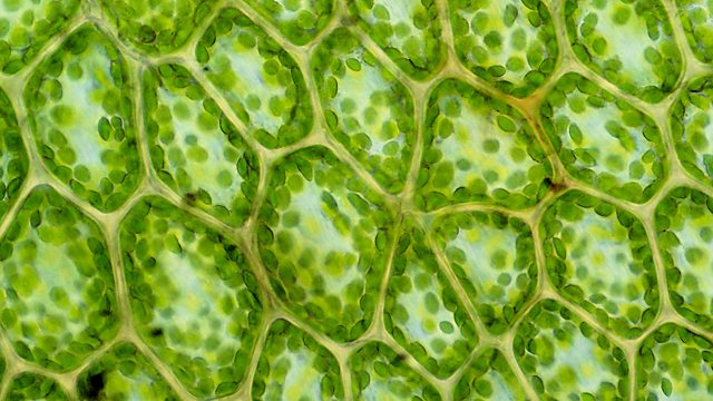
Cells | Create WebQuest from ichef.bbci.co.uk Cells consist of cytoplasm enclosed within a membrane, which contains many biomolecules such as proteins and nucleic acids.2 most plant and animal cells are only visible under a light microscope, with dimensions between 1 and 100 micrometres.3 electron microscopy gives a much higher. Huge collection, amazing choice, 100+ million high quality, affordable rf and rm images. Once slides have been prepared, they can be examined under a microscope. Explanation:the two visible structures that are visible with 'light microscope' are the cell wall and the vacuoles. These organelles are responsible for protein synthesis. Sample has to be placed in a vacuum. This diagram shows a typical animal cell. Describe and compare the structure of a plant cell with an animal cell, as seen under a light microscope, limited to cell wall, nucleus, cytoplasm, chloroplasts, vacuoles and location of the cell membrane.
We say cells are microscopic because they can only be seen under a microscope.
Under a light microscope, the cell membrane, nucleus and cytoplasm of a cheek cell (animal cell) can be observed. A cell is a very tiny structure which exists in living bodies. With a light microscope you can see several structures inside the cell. However, as you probably noticed in the previous activity. It sure will be interesting to see how far they go. Most light microscopes will enlarge a specimen up to 1000 times (1000x) but the electron. Plant cells have cell walls, one large vacuole per cell, and chloroplasts, while animal cells will have a cell membrane only. Detailed images of the cell made with a light microscope generally require staining. Use electromagnets to focus electrons resulting in significantly greater magnifications and resolutions. With a light microscope you can see individual cells and large subcellular structures like the nucleus, but not internal cell structures such as. When you look at animal or plant cells under the electron microscope, you can see a lot more detail. We use microscope comprehensively in microbiology, mineralogy, cell biology, biotechnology, nano physics, microelectronics, pharmacology, and forensics. However, they usually can achieve a maximum of 2000x magnification which is not sufficient to see many other tiny organelles.