Animal Cell Labeled Cilia - Animal Cell Diagram - Labeled - Tim's Printables : (a) control section of the dentate gyrus in a pomc cre;
byAbdul Katzenbach-
0
Animal Cell Labeled Cilia - Animal Cell Diagram - Labeled - Tim's Printables : (a) control section of the dentate gyrus in a pomc cre;. The plural is cilia) is an organelle found on eukaryotic cells in the shape of a slender protuberance that projects from the much larger cell body. Three measurements per cells were averaged. Animal cells are the types of cells that make up most of the tissue cells in animals. The brain of the cell. The main function is to help the cells in terms of movement.
Animals have been evolutionarily successful because of their flexible. The cilium (from latin 'eyelash'; Three measurements per cells were averaged. When the broken cell is damaged beyond recovery it destroys itself, the process is called apoptosis. Some animal cells have cilia or a flagellum.
Animal Cell Diagram Labeled : Biological Science Picture ... from pulpbits.net Animal cell nomenclature book illustrates and describes 19 parts of an animal cell: There are many different types of cells in animals. Cell type specific labeling of cilia using cre mediated activation in the ciliagfpmouse. Primary cilia are vital signaling organelles that extend from most types of cells, including neurons and glia. The animal cell can commit suicide. In contrast, ependymal cells were labeled by brdu administration during embryonic development. In multicellular organisms, cilia function to move fluid or materials past an immobile cell as the cilia does a number of things in the animal cell. Different from other eukaryotic cells, such as plant cells, because they have no cell walls protect cells.
Not all cilia are motile;
Many body cells contain a nonmotile cilium (primary cilium). The cilium of a cell translates varied extracellular cues into intracellular signals that control embryonic development and organ function. There are two types of cilia: The cilium (from latin 'eyelash'; Animal cell, cell membrane, centriole, centrosome, cilia, cytoplasm, cytoskeleton, golgi practice labeling the basic parts of plant and animal cells. Cilia are used to sweep mucus up the airway to prevent infections. The most common examples of ciliated cells are those that line the trachea, or wind pipe, of animals. (a) control section of the dentate gyrus in a pomc cre; There are six animal cell diagrams to choose from. Unlike plants, animals need muscles to move, a nervous system although this image is viewed by light microscopy, the cilia are visible at high magnification. Some eukaryotic cells either have cilia or flagella. Microinject labeled subunits into living cell & they are incorporated into polymeric form of protein, the b. Cell type specific labeling of cilia using cre mediated activation in the ciliagfpmouse.
Microinject labeled subunits into living cell & they are incorporated into polymeric form of protein, the b. Animal cells have a nucleus, organelles, and are surrounded by a cell membrane. Unlike plants, animals need muscles to move, a nervous system although this image is viewed by light microscopy, the cilia are visible at high magnification. The animal cell can commit suicide. The main function is to help the cells in terms of movement.
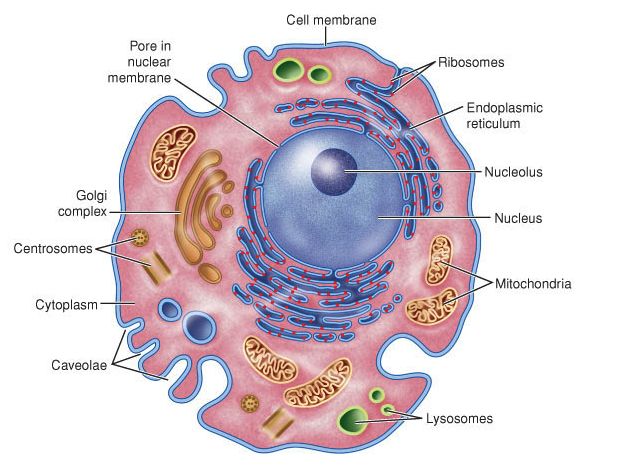
animal cell model labeled, project ideas middle school ... from i.pinimg.com Animal cells are the types of cells that make up most of the tissue cells in animals. It carries information for reproduction and controls all cell activity. The brain of the cell. There are many different types of cells in animals. Animal cell, cell membrane, centriole, centrosome, cilia, cytoplasm, cytoskeleton, golgi practice labeling the basic parts of plant and animal cells. Cilia generally move matter past a cell. Animal cells have a nucleus, organelles, and are surrounded by a cell membrane. These organelles are composed of microtubules, help build flagella and cilia, and appear to be involved in cell division.
The brain of the cell.
(a) control section of the dentate gyrus in a pomc cre; As different markers and fixation were used, the the distribution of the number of centrioles in mature ependymal cells varied between animals from different litters (different sizes) of different genetic. Primary cilia are vital signaling organelles that extend from most types of cells, including neurons and glia. Not all cilia are motile; Animals have been evolutionarily successful because of their flexible. Only present in animal cells and some fungal cells, a pair of centrioles is located near the nucleus, in a region called the centrosome. These are organelles pertinent to plant cells. The cilium (from latin 'eyelash'; In multicellular organisms, cilia function to move fluid or materials past an immobile cell as the cilia does a number of things in the animal cell. Cilia generally move matter past a cell. There, the cilia move mucus containing dirt and other inhaled particles up the windpipe and into the esophagus, where the particles can be coughed up or swallowed. Structure and support for the cell. Role in the formation of flagella, cilia and centrioles.
The cilium (from latin 'eyelash'; Only present in animal cells and some fungal cells, a pair of centrioles is located near the nucleus, in a region called the centrosome. Animals have been evolutionarily successful because of their flexible. Cilia on the surface beat to move fluids and particles up the trachea. Wim cells had ablated cilia and deficiencies in directed migration (electrotaxis), cell proliferation and intracellular signaling.
Labeled Animal Cell - ClipArt Best from www.clipartbest.com Cilia (singular is cilium) are hairlike processes that extend from the cell's surface. These organelles are composed of microtubules, help build flagella and cilia, and appear to be involved in cell division. Notably, protozoans locomote, but it is only via nonmuscular means, in effect, using cilia, flagella, and pseudopodia. The main function of these microscopic organelles is to serve as digestion compartments. The animal cell can commit suicide. As different markers and fixation were used, the the distribution of the number of centrioles in mature ependymal cells varied between animals from different litters (different sizes) of different genetic. (a) control section of the dentate gyrus in a pomc cre; Printable animal cell diagram to help you learn the organelles in an animal cell in preparation for your test or quiz.
(a) control section of the dentate gyrus in a pomc cre;
In this video i'm going to draw labelled diagram of animal cell.in this video you will see the diagram of animal cell and it's labelling.this diagram of. Role in the formation of flagella, cilia and centrioles. Small bubble that carries food/storage. Three measurements per cells were averaged. The main function is to help the cells in terms of movement. The cilium (from latin 'eyelash'; Some eukaryotic cells either have cilia or flagella. Cell type specific labeling of cilia using cre mediated activation in the ciliagfpmouse. The plural is cilia) is an organelle found on eukaryotic cells in the shape of a slender protuberance that projects from the much larger cell body. Olfactory supporting cell microvilli bound only dolichos biflorus agglutinin respiratory cilia bound wga and, somewhat more weakly, pna; Not all cilia are motile; There are many different types of cells in animals. The structure of an animal cell, with labeled parts.