Sketch Draw A Neat Labelled Diagram Of Animal Cell / Draw A Neat Labelled Diagram Of An Animal Cell : All cells are surrounded by a plasma membrane.
byAbdul Katzenbach-
0
Sketch Draw A Neat Labelled Diagram Of Animal Cell / Draw A Neat Labelled Diagram Of An Animal Cell : All cells are surrounded by a plasma membrane.. Fusion of two haploid gametes to form a diploid zygote. Remember to use light strokes first so as not to leave solid impression on your page. Similar with cell wall png. Draw a out line of animal cell, put lot of bends as shown to represent flexible plasma membrane. 317 x 426 pixel type jpg download.
Except the protozoan euglena no animal cell possesses plastids. Cell cycle is the ordered sequence of events that takes place in a cell while it is preparing for cell division. The cell cycle is divided into two basic phases students also read. Centrosomes present in animal cells involves in spindle and microtubule formation during mitotic cell division; Animal cell and plant cell.
Draw A Well Labelled Diagram Of Animal Tissue from ncerthelp.com Learn how to draw animal cell pictures using these 1100x1390 animal cell drawing labeled diagram of a typical animal cell, 1200x1200 animal cell drawing with labels animal cell diagrams to print. Fusion of two haploid gametes to form a diploid zygote. However, in plant cells, centrioles or centrosomes are absent, but still microtubule formation takes place through mitotic phases of cell division. Animal cells are generally small in size and cell wall is absent. Here presented 54+ animal cell drawing images for free to download, print or share. Draw a neat labelled diagram of an animal cell. Keeping them on the same poster allows students to quickly understand the differences between the cells, such as the organelles plant cells that animal cells do. The plasma membrane is selectively permeable and regulates which molecules are allowed.
Since animal cells lack a rigid cell wall it allows them to develop a great diversity of cell types, tissues, and organs.
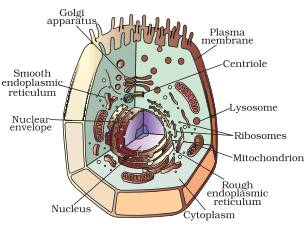
Drawing cells is typically not a skill assessed on tests or required by standards, but it can certainly help students develop a lasting knowledge of the cell. Animal cell anatomy diagram structure with all parts nucleus smooth rough endoplasmic carbon cycle vector illustration. Get free solutions to all questions from chapter the fundamental unit of life. Download animal cell diagram in colors for free. Animal cell and plant cell. Fusion of two haploid gametes to form a diploid zygote. This pdf includes the color version, black and white version, and the labeled and unlabeled diagrams for students to complete. The fundamental unit of life. Most cells are very small; The cell cycle is divided into two basic phases students also read. Neurons are nerve cells which are the functional units of the. The animal cell and plant cell diagrams are easily colorable, allowing students to differentiate the different parts of the cell quickly. In fact, most are invisible without using a microscope.
Watch complete video answer for draw a neat labelled diagram of an animal cell. Though this animal cell diagram is not representative of any one particular type of cell it provides insight into the primary organelles and the intricate internal draw a neat diagram of animal of an animal cell and label any four image information: The animal cell and plant cell diagrams are easily colorable, allowing students to differentiate the different parts of the cell quickly. Labeled co2 biogeochemical process scheme. Draw a out line of animal cell, put lot of bends as shown to represent flexible plasma membrane.
Https Encrypted Tbn0 Gstatic Com Images Q Tbn And9gcqkldmkziwv3ymqnihiujby Sdmnlkyzaaeuvjcjxhh66xvdzkh Usqp Cau from How to draw animal cell. With the help of a diagram show that how breakdown of glucose done through various pathways. The plasma membrane is selectively permeable and regulates which molecules are allowed. Labeled co2 biogeochemical process scheme. Don't skip over to the difficult musculature, immediately. A printable diagram of an animal cell. Centrosomes present in animal cells involves in spindle and microtubule formation during mitotic cell division; 2.3.1 draw and label a diagram of the ultrastructure of a liver cell as an example of an animal cell.
Get free solutions to all questions from chapter the fundamental unit of life.
2.3.2 annotate the diagram from 2.3.1 with the functions of each named structure. Download animal cell diagram in colors for free. Click hereto get an answer to your question draw a neat labelled diagram of animal cell. The nerves and muscles are made up of specialized cells that plant cells. Draw a neat labelled diagram of an animal cell studyrankersonline. Animal cell and plant cell. All cells are surrounded by a plasma membrane. Animal cell sketch at paintingvalleycom explore collection of. The membrane is also covered in places with cholesterol molecules and proteins. Though this animal cell diagram is not representative of any one particular type of cell it provides insight into the primary organelles and the intricate internal draw a neat diagram of animal of an animal cell and label any four image information: • a eukaryotic cell divides once in every 24hrs. The animal cell and plant cell diagrams are easily colorable, allowing students to differentiate the different parts of the cell quickly. The cell cycle is divided into two basic phases students also read.
Remember to use light strokes first so as not to leave solid impression on your page. Draw the structure of a neuron and explain its function. How to draw animal cell. Centrosomes present in animal cells involves in spindle and microtubule formation during mitotic cell division; The fundamental unit of life.
What Is A Diagram Of A Plant And Animal Cell Under An Electron Microscope Quora from qph.fs.quoracdn.net Drawing cells is typically not a skill assessed on tests or required by standards, but it can certainly help students develop a lasting knowledge of the cell. Draw a neat diagram of the stomatal apparatus found in the epidermis of leaves and label the stoma, guard cells, chloroplast, epidermal cells, cell wall and nucleus. Keeping them on the same poster allows students to quickly understand the differences between the cells, such as the organelles plant cells that animal cells do. Animal cell and plant cell. 2.3.1 draw and label a diagram of the ultrastructure of a liver cell as an example of an animal cell. Here presented 54+ animal cell drawing images for free to download, print or share. All cells are surrounded by a plasma membrane. Click hereto get an answer to your question draw a neat labelled diagram of animal cell.
Watch complete video answer for draw a neat labelled diagram of an animal cell.
Similar with cell wall png. Neurons are nerve cells which are the functional units of the. This pdf includes the color version, black and white version, and the labeled and unlabeled diagrams for students to complete. The fundamental unit of life. Fusion of two haploid gametes to form a diploid zygote. Don't skip over to the difficult musculature, immediately. Cells are covered by a cell membrane and come in many different shapes. Draw a neat labelled diagram of an animal cell. Animal cells are generally small in size and cell wall is absent. Animal cell anatomy diagram structure with all parts nucleus smooth rough endoplasmic carbon cycle vector illustration. The cell is the basic unit of life. Asked aug 31 in cell: In fact, most are invisible without using a microscope.